



Congenital urinary tract anomalies are relatively common in pediatric populations. These conditions may lead to urinary tract infections, renal impairment and loss of kidney function; therefore, early detection and timely treatment are essential.
Previously, these anomalies were often detected only after complications had developed; however, in recent years, they are increasingly identified early in the neonatal period, or even prenatally.
Children are commonly brought for evaluation due to symptoms such as dysuria, intermittent or continuous urinary leakage, with urine passing through the normal urethral meatus or ectopic sites such as the vagina. The urine may appear cloudy or pink tinged. In some cases, a palpable mass may be noted in the lower abdomen or flank region.
The following are some of the most common congenital urinary tract anomalies:
Urinary tract anomaly: phimosis
Clinical presentation: dysuria, ballooning of the foreskin during micturition, inability to retract the foreskin, with the urethral meatus not visible.
Management: includes surgical intervention, dilation or manual retraction. In cases of phimosis without a fibrotic ring, gradual retraction can be performed to widen the foreskin. This method is simple, minimally invasive, painless and provides good long term outcomes.
Alternatively, gentle dilation using a small instrument, adhesiolysis and proper cleansing of the glans and coronal sulcus may be performed. However, in the presence of a fibrotic ring or paraphimosis, surgical management is indicated.
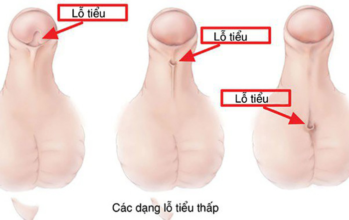
Urethral meatal stenosis
This condition may occur following circumcision, characterized by a narrowed or abnormally positioned urethral meatus.
Clinical presentation: decreased urinary stream and dysuria.
Management: urethral dilation or surgical enlargement of the meatus.
Urethral stricture
This condition may result from trauma, infection or congenital abnormalities. Clinical presentation: dysuria, weak urinary stream and recurrent urinary tract infections. Urethrography is required to determine the length, location and severity of the stricture. Management: depending on the extent of narrowing, treatment options include urethral dilation, endoscopic incision, or surgical resection with urethral reconstruction.
Urethral diverticulum
More common in boys and rare in girls, often presenting soon after birth. Clinical presentation: absence of a normal urinary stream with continuous dribbling, and possible fever due to urinary tract infection. A tense, rounded mass may be palpated in the scrotum; when pressed, urine may be expressed from the urethral meatus. Diagnosis is confirmed by urethrography. Management: antibiotic therapy combined with surgical excision of the diverticulum and urethral reconstruction.
Posterior urethral valves in boys
Clinical presentation: difficulty urinating or continuous dribbling, incomplete bladder emptying leading to bladder distension. Diagnosis is established by imaging studies such as X ray or urethrography. Management: endoscopic valve ablation.
Patent urachus
Clinical presentation: persistent leakage of clear fluid from the umbilicus, either continuously or during urination, with urine passing both through the urethral meatus at the glans penis and through the umbilicus. Diagnosis is confirmed by cystography or by instillation of methylene blue into the bladder or urethra. This condition may resolve spontaneously in neonates; however, persistent cases require surgical excision of the urachal remnant.
Ureterovesical junction obstruction or intramural ureteral stenosis
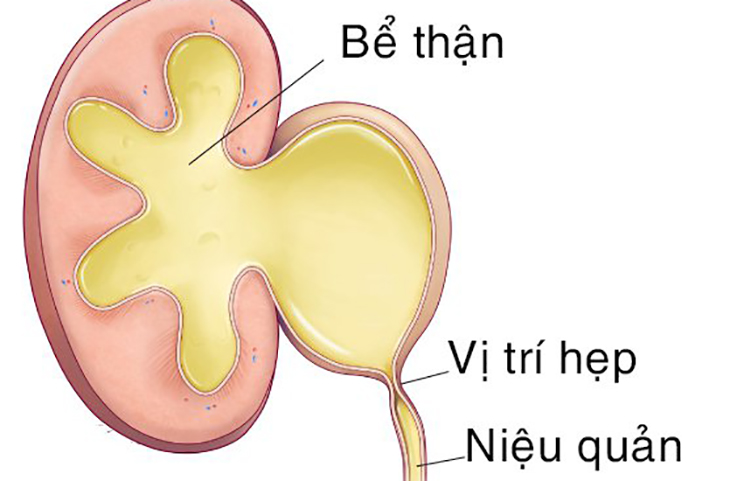
This condition may lead to ureteral dilatation and hydronephrosis. Clinical presentation: cloudy urine, recurrent urinary tract infections, and in some cases, a palpable mass in the flank region. Diagnosis can be established through ultrasonography and radiographic imaging. Management: surgical excision of the stenotic ureteral segment with ureteral reimplantation into the bladder, incorporating an anti reflux mechanism.
Ureteral orifice stenosis
This condition may result in the formation of a ureterocele, which can be located within the bladder or prolapse through the urethral meatus (more commonly in girls), potentially causing urinary retention. Diagnosis is based on ultrasonography, cystography and cystoscopy. Management: endoscopic incision of the ureterocele or surgical excision of the affected ureter and associated poorly functioning renal moiety in cases of duplex systems with significant dilation.
Vesicoureteral reflux
Due to various causes, urine flows retrograde from the bladder into the ureters, leading to ureteral dilatation, urinary stasis, recurrent infections and potential renal impairment. Diagnosis is established by radiography and ultrasonography. Management: mild cases (grades I and II) are treated with antibiotics, while surgical intervention is indicated in more severe cases (grades III and IV).
Ureteral stenosis
Clinical presentation: flank pain, cloudy urine, and a palpable abdominal mass due to renal enlargement. Diagnosis is based on ultrasonography and contrast enhanced imaging of the urinary tract. Management: surgical incision of the obstructive segment, with possible reconstruction of the dilated proximal ureter.
Ureteropelvic junction obstruction
Clinical presentation: flank pain, cloudy urine, and a palpable mass in the flank region due to hydronephrosis. Diagnosis is confirmed by ultrasonography and contrast imaging studies. This anomaly is now often detected prenatally. Management: surgical resection of the narrowed segment, reconstruction of the dilated renal pelvis and reanastomosis of the ureter to the pelvis.
Duplex kidney and ureter
Clinical presentation: duplication of the renal collecting system with two ureters on one or both sides. The ureters may both drain into the bladder, or one may insert ectopically into the urethra, periurethral region or vagina, leading to continuous urinary leakage. Diagnosis is established by ultrasonography and contrast imaging of the urinary tract. Management: surgical intervention is indicated in cases of urinary incontinence or recurrent infections.
Conclusion
Congenital urinary tract anomalies in children are diverse and may coexist or be associated with abnormalities in other organ systems. Early presentation and diagnosis significantly improve treatment outcomes.
In recent months, many pediatric patients have been evaluated and treated based on prenatal diagnoses. Parents are strongly encouraged to seek medical evaluation promptly if any abnormalities are suspected in their children to ensure early intervention.
Reader may also be interested in:
- Intussusception in children: Causes of intussusception
- Pneumonia: Causes of pneumonia in children
- Bronchitis: Common symptoms of bronchitis in children
Note: The information provided in this article by Hong Ngoc General Hospital is for reference purposes only and does not replace professional medical diagnosis or treatment.
For more health related updates, please follow Hong Ngoc General Hospital’s official fanpage: https://www.facebook.com/BenhvienHongNgoc/