Breast fibroadenoma is one of the most common benign breast conditions in women, particularly during the reproductive years. However, in certain cases, fibroadenomas may carry a potential risk of malignant transformation if not closely monitored and appropriately managed.
Join Hong Ngoc General Hospital in exploring the causes, clinical manifestations, diagnostic approaches, and treatment options for this condition.
1. What is breast fibroadenoma? Types of fibroadenoma
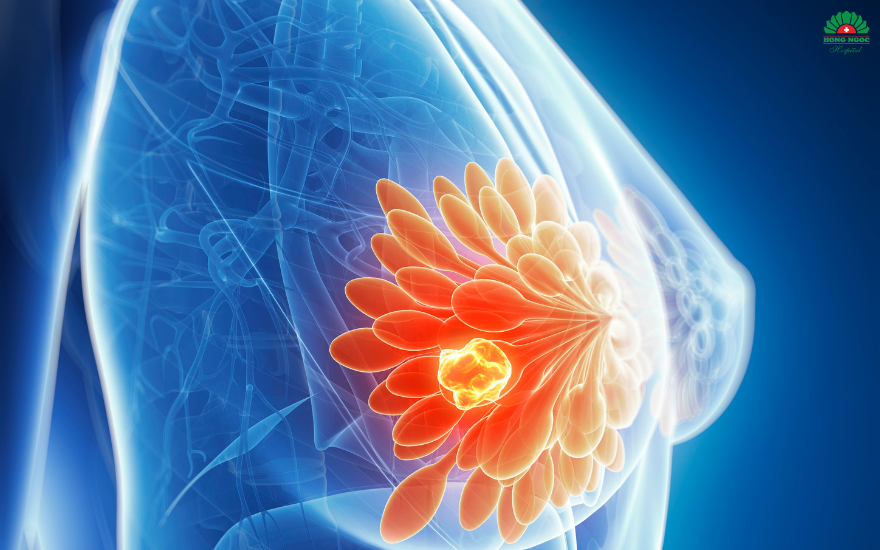
Breast fibroadenoma is a benign tumor that develops within the breast tissue as a result of abnormal proliferation of fibrous and glandular components. The mass is typically round or oval, firm, smooth, and freely mobile beneath the skin, and may occur in one or both breasts.
Based on histopathological characteristics, breast fibroadenomas are classified into four main types:
Simple fibroadenoma: The most common benign form, characterized by a homogeneous structure and well defined margins, with a very low risk of malignant transformation.
Complex fibroadenoma: Associated with additional histological changes such as epithelial hyperplasia, calcifications, or fat necrosis, and carries a relatively higher risk of progression to breast cancer compared with the simple type.
Juvenile fibroadenoma: Typically occurs in adolescent girls due to hormonal fluctuations and may regress spontaneously once hormonal balance is restored.
Giant fibroadenoma: Defined as a fibroadenoma larger than 5 cm in diameter, which may cause breast deformity and often requires surgical excision.
Breast fibroadenomas are tumors that develop within the breast tissue.
2. Common clinical manifestations of breast fibroadenoma
In the early stages, breast fibroadenomas often progress silently, typically without pain or systemic symptoms. However, as the lesion increases in size or responds to hormonal influences, more noticeable signs may develop, including:
A firm, round, and mobile breast mass: This is the most characteristic feature of fibroadenoma. On palpation, the lesion is typically round or oval, well circumscribed, smooth surfaced, and freely movable. The size may range from a few millimeters to several centimeters and can gradually increase over time or become more prominent before menstruation due to hormonal influence.
Breast fullness or mild pain: As the fibroadenoma enlarges or responds to hormonal fluctuations, patients may experience a sensation of heaviness, mild tenderness, or dull discomfort localized to the affected area. Symptoms may intensify with palpation or positional changes.
Changes in breast contour or size: Larger lesions may cause asymmetry, with one breast appearing slightly enlarged. In cases of giant fibroadenoma, noticeable breast deformity may occur, affecting cosmetic appearance.
Abnormal nipple discharge: In rare cases, fibroadenoma may be associated with nipple discharge that is whitish, pale yellow, or blood tinged. This finding warrants further evaluation, as it may indicate atypical hyperplasia or early stage breast carcinoma.
Additional associated signs: Some patients may report itching, skin discoloration, or changes in the periareolar region. Hormonal imbalance may also be accompanied by menstrual irregularities in certain cases.
Note: If the breast mass increases rapidly in size, becomes significantly painful, or is accompanied by skin or nipple changes, prompt evaluation at a qualified healthcare facility is recommended to rule out malignancy.
Breast pain and a palpable firm mass are among the most common clinical manifestations of fibroadenoma.
3. What causes breast fibroadenoma?
The exact etiology of breast fibroadenoma has not been fully established. However, accumulating evidence suggests that hormonal fluctuations, particularly estrogen, play a central role in the development and growth of these lesions.
Several factors are considered to increase the risk of fibroadenoma, including:
3.1. Female hormonal fluctuations
Estrogen and progesterone directly influence the proliferation of breast epithelial and stromal cells. Significant hormonal fluctuations, such as those occurring during the menstrual cycle, puberty, pregnancy, or perimenopause, may stimulate excessive breast tissue growth, contributing to fibroadenoma formation.
Women who use estrogen containing oral contraceptives or undergo long term hormone replacement therapy may have a higher risk of developing breast fibroadenoma compared with the general population.
3.2. Genetic predisposition
A family history of breast fibroadenoma or breast cancer, particularly in first degree relatives such as a mother or sister, may increase individual risk. This association is thought to relate to inherited characteristics of breast tissue structure and hormonal responsiveness.
3.3. Chronic stress and endocrine imbalance
Prolonged stress or sleep deprivation can disrupt the hypothalamic–pituitary–ovarian axis, leading to hormonal imbalance. Altered regulation of sex hormones may contribute to abnormal breast tissue proliferation and fibroadenoma development.
3.4. Diet and lifestyle factors
High intake of saturated fats, red meat, processed foods, and alcohol may elevate circulating estrogen levels. In addition, a sedentary lifestyle, overweight, and obesity, particularly central obesity, have been associated with an increased risk of breast fibroadenoma.
Chronic stress may increase the risk of developing breast fibroadenoma by disrupting hormonal balance.
4. Is breast fibroadenoma dangerous?
The majority of breast fibroadenomas are benign and do not pose a direct threat to life, nor do they typically affect fertility or breastfeeding. However, a small proportion of cases may carry an increased risk of malignant transformation, particularly complex fibroadenomas or those associated with atypical hyperplasia.
If not appropriately monitored and managed, breast fibroadenomas may lead to the following complications:
Breast deformity: Some fibroadenomas initially measure only a few millimeters but may gradually enlarge to several centimeters, occasionally occupying a significant portion of the breast tissue and causing visible asymmetry or deformity.
Persistent pain: As the mass increases in size and compresses adjacent glandular tissue, patients may experience localized breast pain or tenderness, which can worsen with palpation or positional changes.
Recurrence or multifocal lesions: Despite medical or surgical treatment, fibroadenomas may recur, particularly in women with ongoing hormonal imbalance or unmodified lifestyle factors. In some cases, multiple lesions may develop in one or both breasts, complicating surveillance and management.
Malignant transformation: Complex fibroadenomas are associated with a higher risk of progression to breast carcinoma. This risk is more pronounced in women over 35 years of age, those with a family history of breast cancer, or individuals with chronic hormonal disorders.
Psychological impact and reduced quality of life: The detection of a breast mass may cause significant anxiety, sleep disturbances, and fear of cancer. Large or deforming lesions may also affect body image and self confidence.
Complex fibroadenomas may carry a risk of progression to breast cancer, potentially affecting overall health if not appropriately monitored.
5. Diagnostic approaches for breast fibroadenoma
Most breast fibroadenomas grow slowly and are often painless. As a result, many women only become aware of the condition when it begins to affect daily activities or overall health.
Specialists recommend monthly breast self examination, ideally performed 5–7 days after menstruation, along with annual clinical evaluation at a reputable healthcare facility. Modern diagnostic modalities include:
Clinical breast examination: The physician performs palpation to evaluate the size, consistency, mobility, and margins of the lesion.
Breast ultrasound: The most commonly utilized imaging modality, capable of detecting small fibroadenomas and providing detailed assessment of lesion morphology, size, and internal architecture.
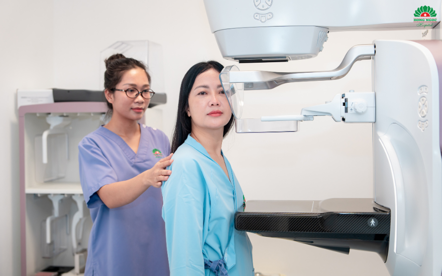
Mammography: Generally indicated for women aged 40 years and older or when a complex lesion is suspected. Mammography is effective in detecting microcalcifications, which may represent early signs of breast carcinoma.
Fine needle aspiration (FNA) or core needle biopsy (CNB): If ultrasound findings are inconclusive, a tissue sample is obtained for cytological or histopathological analysis.
Vacuum assisted breast biopsy (VABB): Performed under ultrasound guidance, this technique enables targeted tissue sampling and, in selected cases, complete lesion removal. It is a critical step in confirming the diagnosis and excluding malignancy.
Mammography helps detect small calcified lesions, including microcalcifications.
6. Management of breast fibroadenoma
Whether surgical intervention is required depends on the lesion’s characteristics, size, clinical symptoms, and biopsy findings. The three most commonly applied management approaches include:
6.1. Periodic surveillance
For small, asymptomatic, and slow growing fibroadenomas, physicians may recommend regular follow up with clinical breast examination and ultrasound monitoring. In some cases, lesions may regress spontaneously once hormonal levels stabilize or after menopause.
6.2. Medical management
In patients with hormonal imbalance or cyclical mastalgia, treatment options may include:
Hormonal regulatory therapy under close medical supervision
Mild analgesics or anti inflammatory agents
Supplementation with vitamin E and omega-3 fatty acids, along with reduced caffeine intake to minimize breast tissue stimulation.
It is important to maintain emotional stability, ensure adequate sleep, and keep a healthy body weight to support hormonal balance, thereby reducing the risk of rapid fibroadenoma growth.
Hormonal therapy may be prescribed to help slow the growth of breast fibroadenoma, under appropriate medical supervision.
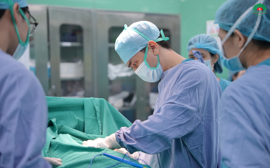
6.3. Surgical excision of breast fibroadenoma
Surgical removal may be indicated when the lesion is large, demonstrates rapid growth, causes breast deformity, or is associated with persistent pain. Excision is particularly recommended if biopsy findings reveal a complex fibroadenoma or features suspicious for malignancy.
With advances in surgical techniques, many hospitals, including Hong Ngoc General Hospital, offer breast conserving excision procedures or vacuum assisted breast biopsy (VABB) for complete lesion removal. These approaches help ensure effective treatment while minimizing scarring and preserving cosmetic outcomes.
Surgical intervention is indicated in cases of large breast tumors associated with complications or suspected malignant potential.
7. Prevention of breast fibroadenoma
Breast fibroadenoma is a benign condition closely associated with female hormonal balance and lifestyle factors. Therefore, women can adopt the following preventive measures to reduce the risk of new lesion development:
Maintain a healthy weight and balanced diet: A diet rich in green vegetables, fresh fruits, whole grains, nuts, and omega-3–rich fish, while limiting fried foods, refined sugars, fast food, and red meat, may help reduce oxidative stress in breast tissue. Adequate hydration is also recommended.
Limit alcohol, caffeine, and stimulants: Evidence suggests that caffeine, alcohol, and tobacco may influence estrogen activity and potentially increase the risk of fibroadenoma development. Women are advised to limit caffeine intake to less than one cup per day, avoid regular alcohol consumption, and refrain from smoking or exposure to secondhand smoke.
Engage in regular physical activity: Moderate exercise such as brisk walking, yoga, swimming, or cycling for at least 30 minutes daily can improve circulation and hormonal metabolism, support weight control, and reduce excess estrogen levels, thereby lowering the risk of fibroadenoma progression.
Manage stress and ensure adequate sleep: Maintaining regular sleep patterns (7–8 hours per night) and practicing stress reduction strategies help stabilize hormonal balance and may reduce the risk of tumor growth or recurrence after treatment.
Routine breast examination and screening: Regular screening is the most effective preventive measure. Women aged 20–40 years are advised to undergo clinical breast examination every 6–12 months. Women over 40 years should combine clinical evaluation with mammography to detect early microcalcifications or small lesions.
Use hormonal therapy only under medical indication: Oral contraceptives, hormone therapy, or exogenous estrogen should not be used without professional guidance, as inappropriate use may stimulate tumor growth.
Light to moderate exercise helps reduce stress and improve hormonal balance, thereby supporting the prevention of breast fibroadenoma.
8. Breast fibroadenoma diagnosis and treatment at Hong Ngoc General Hospital – safe and reliable care
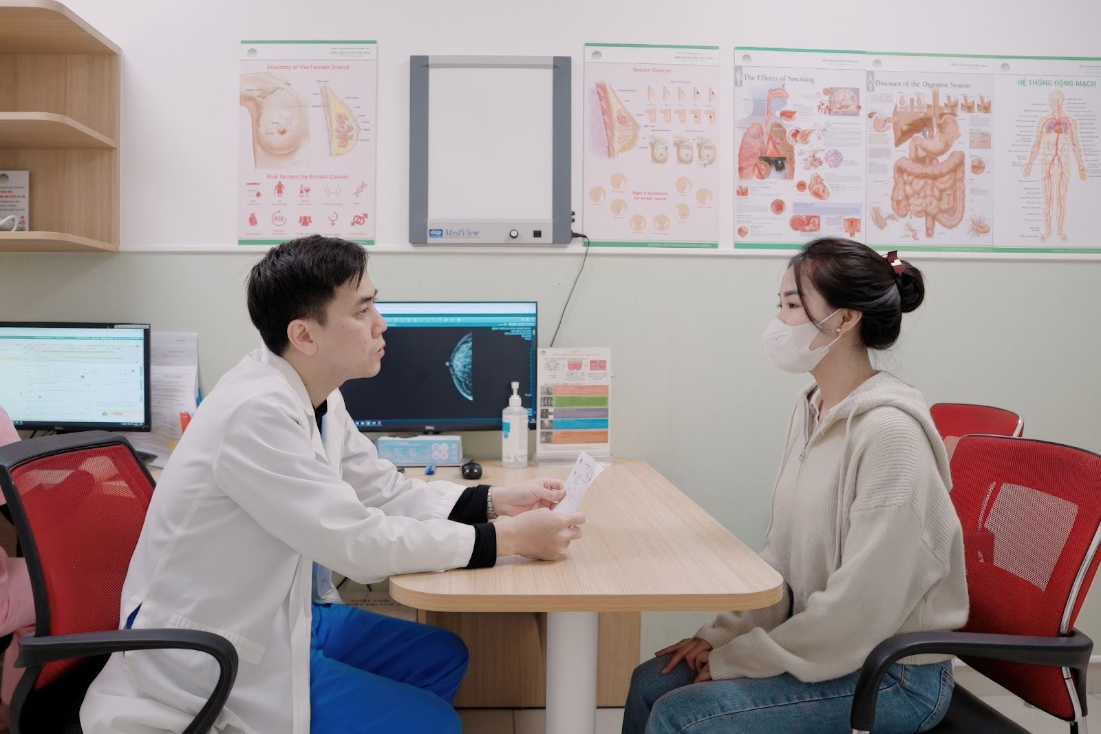
With over 20 years of experience in comprehensive women’s healthcare, Hong Ngoc General Hospital is recognized as a trusted provider in the examination, diagnosis, and management of breast conditions, including fibroadenoma.
At Hong Ngoc, patients are evaluated directly by experienced specialists in obstetrics and gynecology and oncology, many of whom have worked at leading tertiary hospitals nationwide. In addition to strong clinical expertise, the medical team provides thorough consultation, ensuring patients clearly understand their condition and receive individualized, evidence based treatment recommendations.
The hospital is equipped with advanced medical technology, including:
High resolution breast ultrasound systems integrated with AI technology, such as Voluson Expert 22 and Logiq Fortis, enabling automated detection and measurement of small lesions, as well as guidance for biopsy in cases suspicious for malignancy.
Digital mammography systems, providing high clarity imaging and supporting early detection of microcalcifications.
Vacuum assisted breast biopsy (VABB), a minimally invasive technique for the removal of benign breast lesions without open surgery. This approach preserves breast aesthetics, promotes faster recovery, and minimizes post procedural pain and scarring.
Regular breast examination and screening help detect fibroadenoma at an early stage and enable timely management.
Contact 091 669 0018 or visit the official fanpage of Hong Ngoc General Hospital to schedule your appointment today.
Note: The information provided in this article by Hong Ngoc General Hospital is for reference only and does not replace professional medical diagnosis or treatment. Patients should not self medicate. For an accurate assessment of their condition, individuals are advised to visit a qualified healthcare facility for direct examination, diagnosis, and appropriate treatment consultation by a physician.
Breast fibroadenoma is one of the most common benign breast conditions in women, particularly during the reproductive years. However, in certain cases, fibroadenomas may carry a potential risk of malignant transformation if not closely monitored and appropriately managed.
Join Hong Ngoc General Hospital in exploring the causes, clinical manifestations, diagnostic approaches, and treatment options for this condition.
1. What is breast fibroadenoma? Types of fibroadenoma
Breast fibroadenoma is a benign tumor that develops within the breast tissue as a result of abnormal proliferation of fibrous and glandular components. The mass is typically round or oval, firm, smooth, and freely mobile beneath the skin, and may occur in one or both breasts.
Based on histopathological characteristics, breast fibroadenomas are classified into four main types:
Simple fibroadenoma: The most common benign form, characterized by a homogeneous structure and well defined margins, with a very low risk of malignant transformation.
Complex fibroadenoma: Associated with additional histological changes such as epithelial hyperplasia, calcifications, or fat necrosis, and carries a relatively higher risk of progression to breast cancer compared with the simple type.
Juvenile fibroadenoma: Typically occurs in adolescent girls due to hormonal fluctuations and may regress spontaneously once hormonal balance is restored.
Giant fibroadenoma: Defined as a fibroadenoma larger than 5 cm in diameter, which may cause breast deformity and often requires surgical excision.
Breast fibroadenomas are tumors that develop within the breast tissue.
2. Common clinical manifestations of breast fibroadenoma
In the early stages, breast fibroadenomas often progress silently, typically without pain or systemic symptoms. However, as the lesion increases in size or responds to hormonal influences, more noticeable signs may develop, including:
A firm, round, and mobile breast mass: This is the most characteristic feature of fibroadenoma. On palpation, the lesion is typically round or oval, well circumscribed, smooth surfaced, and freely movable. The size may range from a few millimeters to several centimeters and can gradually increase over time or become more prominent before menstruation due to hormonal influence.
Breast fullness or mild pain: As the fibroadenoma enlarges or responds to hormonal fluctuations, patients may experience a sensation of heaviness, mild tenderness, or dull discomfort localized to the affected area. Symptoms may intensify with palpation or positional changes.
Changes in breast contour or size: Larger lesions may cause asymmetry, with one breast appearing slightly enlarged. In cases of giant fibroadenoma, noticeable breast deformity may occur, affecting cosmetic appearance.
Abnormal nipple discharge: In rare cases, fibroadenoma may be associated with nipple discharge that is whitish, pale yellow, or blood tinged. This finding warrants further evaluation, as it may indicate atypical hyperplasia or early stage breast carcinoma.
Additional associated signs: Some patients may report itching, skin discoloration, or changes in the periareolar region. Hormonal imbalance may also be accompanied by menstrual irregularities in certain cases.
Note: If the breast mass increases rapidly in size, becomes significantly painful, or is accompanied by skin or nipple changes, prompt evaluation at a qualified healthcare facility is recommended to rule out malignancy.
Breast pain and a palpable firm mass are among the most common clinical manifestations of fibroadenoma.
3. What causes breast fibroadenoma?
The exact etiology of breast fibroadenoma has not been fully established. However, accumulating evidence suggests that hormonal fluctuations, particularly estrogen, play a central role in the development and growth of these lesions.
Several factors are considered to increase the risk of fibroadenoma, including:
3.1. Female hormonal fluctuations
Estrogen and progesterone directly influence the proliferation of breast epithelial and stromal cells. Significant hormonal fluctuations, such as those occurring during the menstrual cycle, puberty, pregnancy, or perimenopause, may stimulate excessive breast tissue growth, contributing to fibroadenoma formation.
Women who use estrogen containing oral contraceptives or undergo long term hormone replacement therapy may have a higher risk of developing breast fibroadenoma compared with the general population.
3.2. Genetic predisposition
A family history of breast fibroadenoma or breast cancer, particularly in first degree relatives such as a mother or sister, may increase individual risk. This association is thought to relate to inherited characteristics of breast tissue structure and hormonal responsiveness.
3.3. Chronic stress and endocrine imbalance
Prolonged stress or sleep deprivation can disrupt the hypothalamic–pituitary–ovarian axis, leading to hormonal imbalance. Altered regulation of sex hormones may contribute to abnormal breast tissue proliferation and fibroadenoma development.
3.4. Diet and lifestyle factors
High intake of saturated fats, red meat, processed foods, and alcohol may elevate circulating estrogen levels. In addition, a sedentary lifestyle, overweight, and obesity, particularly central obesity, have been associated with an increased risk of breast fibroadenoma.
Chronic stress may increase the risk of developing breast fibroadenoma by disrupting hormonal balance.
4. Is breast fibroadenoma dangerous?
The majority of breast fibroadenomas are benign and do not pose a direct threat to life, nor do they typically affect fertility or breastfeeding. However, a small proportion of cases may carry an increased risk of malignant transformation, particularly complex fibroadenomas or those associated with atypical hyperplasia.
If not appropriately monitored and managed, breast fibroadenomas may lead to the following complications:
Breast deformity: Some fibroadenomas initially measure only a few millimeters but may gradually enlarge to several centimeters, occasionally occupying a significant portion of the breast tissue and causing visible asymmetry or deformity.
Persistent pain: As the mass increases in size and compresses adjacent glandular tissue, patients may experience localized breast pain or tenderness, which can worsen with palpation or positional changes.
Recurrence or multifocal lesions: Despite medical or surgical treatment, fibroadenomas may recur, particularly in women with ongoing hormonal imbalance or unmodified lifestyle factors. In some cases, multiple lesions may develop in one or both breasts, complicating surveillance and management.
Malignant transformation: Complex fibroadenomas are associated with a higher risk of progression to breast carcinoma. This risk is more pronounced in women over 35 years of age, those with a family history of breast cancer, or individuals with chronic hormonal disorders.
Psychological impact and reduced quality of life: The detection of a breast mass may cause significant anxiety, sleep disturbances, and fear of cancer. Large or deforming lesions may also affect body image and self confidence.
Complex fibroadenomas may carry a risk of progression to breast cancer, potentially affecting overall health if not appropriately monitored.
5. Diagnostic approaches for breast fibroadenoma
Most breast fibroadenomas grow slowly and are often painless. As a result, many women only become aware of the condition when it begins to affect daily activities or overall health.
Specialists recommend monthly breast self examination, ideally performed 5–7 days after menstruation, along with annual clinical evaluation at a reputable healthcare facility. Modern diagnostic modalities include:
Clinical breast examination: The physician performs palpation to evaluate the size, consistency, mobility, and margins of the lesion.
Breast ultrasound: The most commonly utilized imaging modality, capable of detecting small fibroadenomas and providing detailed assessment of lesion morphology, size, and internal architecture.
Mammography: Generally indicated for women aged 40 years and older or when a complex lesion is suspected. Mammography is effective in detecting microcalcifications, which may represent early signs of breast carcinoma.
Fine needle aspiration (FNA) or core needle biopsy (CNB): If ultrasound findings are inconclusive, a tissue sample is obtained for cytological or histopathological analysis.
Vacuum assisted breast biopsy (VABB): Performed under ultrasound guidance, this technique enables targeted tissue sampling and, in selected cases, complete lesion removal. It is a critical step in confirming the diagnosis and excluding malignancy.
Mammography helps detect small calcified lesions, including microcalcifications.
6. Management of breast fibroadenoma
Whether surgical intervention is required depends on the lesion’s characteristics, size, clinical symptoms, and biopsy findings. The three most commonly applied management approaches include:
6.1. Periodic surveillance
For small, asymptomatic, and slow growing fibroadenomas, physicians may recommend regular follow up with clinical breast examination and ultrasound monitoring. In some cases, lesions may regress spontaneously once hormonal levels stabilize or after menopause.
6.2. Medical management
In patients with hormonal imbalance or cyclical mastalgia, treatment options may include:
Hormonal regulatory therapy under close medical supervision
Mild analgesics or anti inflammatory agents
Supplementation with vitamin E and omega-3 fatty acids, along with reduced caffeine intake to minimize breast tissue stimulation.
It is important to maintain emotional stability, ensure adequate sleep, and keep a healthy body weight to support hormonal balance, thereby reducing the risk of rapid fibroadenoma growth.
Hormonal therapy may be prescribed to help slow the growth of breast fibroadenoma, under appropriate medical supervision.
6.3. Surgical excision of breast fibroadenoma
Surgical removal may be indicated when the lesion is large, demonstrates rapid growth, causes breast deformity, or is associated with persistent pain. Excision is particularly recommended if biopsy findings reveal a complex fibroadenoma or features suspicious for malignancy.
With advances in surgical techniques, many hospitals, including Hong Ngoc General Hospital, offer breast conserving excision procedures or vacuum assisted breast biopsy (VABB) for complete lesion removal. These approaches help ensure effective treatment while minimizing scarring and preserving cosmetic outcomes.
Surgical intervention is indicated in cases of large breast tumors associated with complications or suspected malignant potential.
7. Prevention of breast fibroadenoma
Breast fibroadenoma is a benign condition closely associated with female hormonal balance and lifestyle factors. Therefore, women can adopt the following preventive measures to reduce the risk of new lesion development:
Maintain a healthy weight and balanced diet: A diet rich in green vegetables, fresh fruits, whole grains, nuts, and omega-3–rich fish, while limiting fried foods, refined sugars, fast food, and red meat, may help reduce oxidative stress in breast tissue. Adequate hydration is also recommended.
Limit alcohol, caffeine, and stimulants: Evidence suggests that caffeine, alcohol, and tobacco may influence estrogen activity and potentially increase the risk of fibroadenoma development. Women are advised to limit caffeine intake to less than one cup per day, avoid regular alcohol consumption, and refrain from smoking or exposure to secondhand smoke.
Engage in regular physical activity: Moderate exercise such as brisk walking, yoga, swimming, or cycling for at least 30 minutes daily can improve circulation and hormonal metabolism, support weight control, and reduce excess estrogen levels, thereby lowering the risk of fibroadenoma progression.
Manage stress and ensure adequate sleep: Maintaining regular sleep patterns (7–8 hours per night) and practicing stress reduction strategies help stabilize hormonal balance and may reduce the risk of tumor growth or recurrence after treatment.
Routine breast examination and screening: Regular screening is the most effective preventive measure. Women aged 20–40 years are advised to undergo clinical breast examination every 6–12 months. Women over 40 years should combine clinical evaluation with mammography to detect early microcalcifications or small lesions.
Use hormonal therapy only under medical indication: Oral contraceptives, hormone therapy, or exogenous estrogen should not be used without professional guidance, as inappropriate use may stimulate tumor growth.
Light to moderate exercise helps reduce stress and improve hormonal balance, thereby supporting the prevention of breast fibroadenoma.
8. Breast fibroadenoma diagnosis and treatment at Hong Ngoc General Hospital – safe and reliable care
With over 20 years of experience in comprehensive women’s healthcare, Hong Ngoc General Hospital is recognized as a trusted provider in the examination, diagnosis, and management of breast conditions, including fibroadenoma.
At Hong Ngoc, patients are evaluated directly by experienced specialists in obstetrics and gynecology and oncology, many of whom have worked at leading tertiary hospitals nationwide. In addition to strong clinical expertise, the medical team provides thorough consultation, ensuring patients clearly understand their condition and receive individualized, evidence based treatment recommendations.
The hospital is equipped with advanced medical technology, including:
High resolution breast ultrasound systems integrated with AI technology, such as Voluson Expert 22 and Logiq Fortis, enabling automated detection and measurement of small lesions, as well as guidance for biopsy in cases suspicious for malignancy.
Digital mammography systems, providing high clarity imaging and supporting early detection of microcalcifications.
Vacuum assisted breast biopsy (VABB), a minimally invasive technique for the removal of benign breast lesions without open surgery. This approach preserves breast aesthetics, promotes faster recovery, and minimizes post procedural pain and scarring.
Regular breast examination and screening help detect fibroadenoma at an early stage and enable timely management.
Contact 091 669 0018 or visit the official fanpage of Hong Ngoc General Hospital to schedule your appointment today.
Note: The information provided in this article by Hong Ngoc General Hospital is for reference only and does not replace professional medical diagnosis or treatment. Patients should not self medicate. For an accurate assessment of their condition, individuals are advised to visit a qualified healthcare facility for direct examination, diagnosis, and appropriate treatment consultation by a physician.
Để lại câu hỏi của bạn để nhận được giải đáp từ các bác sĩ của Hồng Ngọc
Lorem ipsum dolor
Lorem ipsum dolor sit amet, consectetur adipiscing elit. Mauris odio lectus, pretium faucibus nisi eu, accumsan consectetur orci. In blandit vehicula nisl, vel lacinia ligula finibus a. Donec fermentum rhoncus
Lorem ipsum dolor sit amet, consectetur adipiscing elit. Mauris odio lectus, pretium faucibus nisi eu, accumsan consectetur orci. In blandit vehicula nisl, vel lacinia ligula finibus a. Donec fermentum rhoncus
Lorem ipsum dolor sit amet, consectetur adipiscing elit. Mauris odio lectus, pretium faucibus nisi eu, accumsan consectetur orci. In blandit vehicula nisl, vel lacinia ligula finibus a. Donec fermentum rhoncus
Lorem ipsum dolor sit amet, consectetur adipiscing elit. Mauris odio lectus, pretium faucibus nisi eu, accumsan consectetur orci. In blandit vehicula nisl, vel lacinia ligula finibus a. Donec fermentum rhoncus
Lorem ipsum dolor sit amet, consectetur adipiscing elit. Mauris odio lectus, pretium faucibus nisi eu, accumsan consectetur orci. In blandit vehicula nisl, vel lacinia ligula finibus a. Donec fermentum rhoncus