


An 18-year-old male student with advanced keratoconus underwent pinhole pupilloplasty combined with intraocular lens implantation at Hong Ngoc General Hospital, successfully restoring vision without the need for corneal transplantation and eliminating the subsequent risk of graft rejection.

In this situation, corneal transplantation is often considered one of the standard treatment options when the damaged cornea no longer responds adequately to conservative approaches. However, drawing on her extensive experience in ophthalmic surgery, Dinh Thi Hoang Anh, MD, PhD, carefully reassessed the case before proceeding with transplantation.
According to Dr. Hoang Anh, younger patients may face a higher risk of corneal graft rejection because of a more active immune response. If rejection occurs, prolonged treatment may be required and the surgical outcome can be significantly affected. After an individualized assessment of N.’s condition, the team found that although the corneal scar was relatively large and located near the central cornea, it did not completely involve the visual axis. A portion of the cornea remained relatively clear and unobstructed by scar tissue.
These two considerations formed the basis for a different, yet clinically well-founded treatment decision: pinhole pupilloplasty for advanced keratoconus.
Pinhole pupilloplasty: a new cornea-preserving treatment approach for patients with keratoconus
Pinhole pupilloplasty is an advanced ophthalmic surgical technique designed to reconstruct the shape, size, and position of the pupil - the aperture that regulates the amount of light entering the eye. The technique has traditionally been used to manage complex iris injuries, such as iris tears, iridodialysis, or loss of normal iris support.
However, using pinhole pupilloplasty to restore the optical pathway in a patient with advanced keratoconus represents a different therapeutic approach. When the cornea becomes severely distorted, scarred, and associated with profound visual impairment, corneal transplantation is often considered the conventional option. Yet in a young patient who still retains areas of relatively clear cornea, the ophthalmic team at Hong Ngoc General Hospital asked a different question: Could the remaining healthy corneal tissue be utilized to improve vision, rather than replacing the entire cornea with donor tissue?
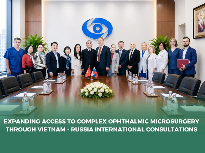
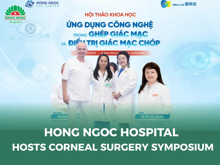
With the goal of preserving as much of the eye’s natural anatomy as possible, Dinh Thi Hoang Anh, MD, PhD, and her team explored an alternative treatment strategy rather than proceeding directly to corneal transplantation. The case underwent an in-depth multidisciplinary consultation with Prof. Kalinnikov Yury, MD, PhD, Founder and General Director of AMG Eye Hospital in Moscow and a leading ophthalmic specialist in the Russian Federation. Specialists from both countries jointly evaluated the corneal architecture, location of the scar, visual axis, and remaining clear corneal tissue to determine the most appropriate individualized treatment plan.
Following the consultation, the team agreed to perform pinhole pupilloplasty, making use of the patient’s remaining transparent cornea instead of replacing it with donor tissue. This decision also reflects the individualized treatment philosophy at the Hong Ngoc Eye Center: not only addressing the current lesion, but also considering long-term preservation of ocular structures, visual function, and overall patient benefit.
The principle of the technique can be understood as creating a new “optical window” for light to enter the eye. The surgeon repositions and reshapes the pupil so that it avoids the scarred corneal area while remaining aligned with the visual axis. As a result, instead of being obstructed by the scar, incoming light is directed through the clearer portion of the cornea, allowing it to enter the eye and form a more functional retinal image.
Rather than “replacing” the cornea, the surgical team chose to preserve and make use of the tissue the patient still had. This approach enabled N. to retain his own cornea, improve visual function, and avoid immediate dependence on donor corneal tissue - an especially meaningful advantage for a young patient.

Although the surgical concept is theoretically straightforward, its clinical execution requires an exceptionally high level of precision. “The surgeon must create a pupil measuring only 1.0–1.5 mm and position it exactly at the preoperatively determined location. Every microsurgical maneuver is performed within the confined space of the eye, where even a minor deviation can damage the iris or result in an irregular, decentered pupil,” explained Dinh Thi Hoang Anh, MD, PhD.
To address this technical challenge, Dr. Hoang Anh used a second-generation smart pupil-sizing device, which she co-invented and developed with a Russian ophthalmic specialist.
The device enables direct measurement at the pupillary plane, rather than relying on estimation through the corneal plane, while also assisting with suture grasping and traction during pupilloplasty. This provides an important basis for precise control of the pupil’s postoperative size, shape, and position.
During the same procedure, the surgical team also performed phacoemulsification with intraocular lens implantation to simultaneously address the patient’s cataract.