


Benign lung tumours are noncancerous lesions that do not metastasise and generally do not pose an immediate threat to life. The most common type is pulmonary hamartoma. Most are discovered incidentally during routine chest X rays or CT scans because they cause no obvious symptoms. However, larger tumours may compress the airways, cause recurrent infections and need to be carefully distinguished from early stage lung cancer.
What is a benign lung tumour?
A benign lung tumour is an abnormal growth of cells in the lung, bronchial or vascular tissue. Unlike malignant tumours, it does not invade surrounding tissues or spread to other organs.
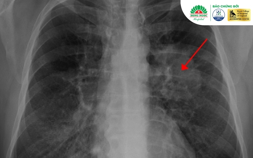
Not every mass or nodule detected on lung imaging can be immediately identified as benign. Doctors assess its imaging characteristics and changes over time. In some cases, histopathological examination is required to determine the nature of the lesion.
Are benign lung tumours dangerous?
Benign lung tumours generally do not pose the same immediate threat to life as lung cancer. However, they should not be overlooked, as they may cause health complications if not appropriately monitored:
- Airway compression: A large tumour may compress or obstruct a bronchus, narrowing the airway, reducing ventilation and potentially causing lung collapse.
- Recurrent infections: Bronchial obstruction may cause secretions to accumulate, increasing the risk of recurrent pneumonia or persistent lung abscesses.
- Bleeding: Some highly vascular tumours may bleed, potentially causing significant haemoptysis.
- Difficulty distinguishing them from malignant lesions: Some benign lung tumours have imaging features similar to those of malignant lesions. Doctors may therefore recommend imaging surveillance, additional diagnostic tests or tissue sampling to determine the nature of the lesion.
Common types of benign lung tumours
Benign lung tumours may arise from different types of tissue within the chest. Common examples include:
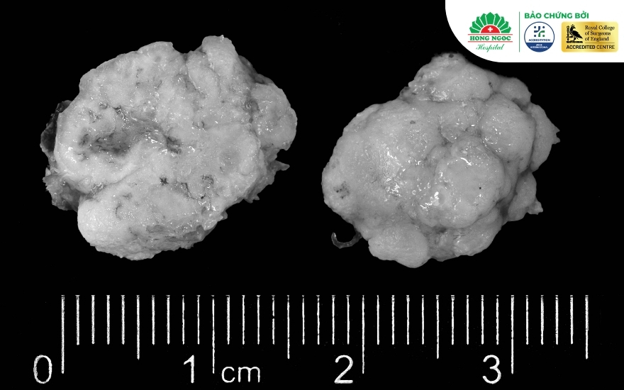
- Pulmonary hamartoma: This is the most common benign lung tumour. It consists of an abnormal mixture of tissues such as cartilage, fat and glandular tissue. These tumours are usually located near the outer part of the lung, grow slowly and have an extremely low risk of malignant transformation.
- Bronchial adenoma: This is a historical term used for several tumours arising from the bronchial glands. Some tumours previously classified under this term, including bronchial carcinoid tumours, are now recognised as having malignant potential and require careful assessment. They may obstruct the bronchial lumen and cause symptoms such as coughing or haemoptysis.
- Other benign tumours:
- Leiomyoma: Arises from bronchial smooth muscle.
- Fibroma and lipoma: Develop from connective tissue or fatty tissue beneath the bronchial lining.
- Schwannoma and neurofibroma: More commonly occur in the mediastinum but may occasionally arise within the lung.
- Benign fibrous histiocytoma: A rare tumour that may develop in the lung.
- Haemangioma: Develops from abnormal blood vessels within the lung.
Signs and symptoms of benign lung tumours
Most benign lung tumours cause no symptoms in their early stages and are discovered incidentally during routine chest X rays or CT scans. When symptoms do occur, they vary according to the location of the tumour, whether in a central bronchus or the peripheral lung tissue, and its size.
Incidental detection on chest X ray or CT
Most benign lung tumours do not cause noticeable symptoms. In the early stages, the tumour is often small, located in the peripheral lung tissue and does not compress the bronchi or pleura.
It is therefore commonly discovered by chance during a routine health check or when a chest X ray or CT scan is performed for another reason.
Warning signs as the tumour grows
If a benign lung tumour grows within or compresses a major bronchus, patients may experience:
- Persistent cough: A dry or productive cough that does not improve with conventional medical treatment.
- Coughing up blood: Blood streaks in the sputum caused by damage to the bronchial lining or small blood vessels. This may occur with certain bronchial tumours or vascular malformations.
- Localised wheezing or shortness of breath: Caused by narrowing or obstruction of the airway.
- Chest pain or discomfort: A dull pain that may worsen when taking a deep breath or coughing.
- Recurrent respiratory infections: Repeated episodes of pneumonia or bronchitis affecting the same lobe or segment of the lung due to impaired drainage of mucus caused by airway obstruction.
Distinguishing benign lung tumours from malignant tumours
Because benign lung tumours and lung cancer may share similar symptoms in their early stages, accurate differentiation is essential for determining the appropriate treatment plan.
CRITERIA | BENIGN LUNG TUMOUR | MALIGNANT LUNG TUMOUR (LUNG CANCER) |
| Cellular characteristics | Well differentiated cells without invasive features | Malignant cellular changes, cellular atypia and abnormal mitotic activity |
| Growth rate | Usually grows very slowly or remains stable for years | May grow rapidly and increase in size over a relatively short period |
| Invasive potential | Does not invade surrounding tissues | May invade the bronchi, pleura or chest wall and spread to the mediastinal lymph nodes, brain, bones, liver or adrenal glands |
| CT features | Smooth, rounded margins with well defined borders; may contain popcorn calcification | Irregular or spiculated margins with poorly defined borders and possible signs of local invasion |
| General symptoms | Usually has little effect on overall health and does not cause sudden weight loss | May be associated with rapid weight loss, fatigue, enlarged lymph nodes and persistent low grade fever |
Note: Findings on chest X rays or CT scans are suggestive only. To determine the nature of the tumour accurately, a thoracic specialist may recommend additional diagnostic procedures such as CT guided lung biopsy, bronchoscopy or histopathological examination.
Treatment of benign lung tumours: When is surgery needed?
Not every benign lung tumour requires immediate surgery. The appropriate treatment depends on the tumour’s size, location, characteristics and associated risks.
Regular monitoring
For small lesions without suspicious features in patients at low risk, doctors may recommend monitoring with follow up CT scans. The timing of surveillance is individualised according to the lesion’s size, imaging characteristics and type, as well as the patient’s risk factors.
Medical treatment of the underlying cause
Some noncancerous conditions may resemble lung tumours and require treatment of the underlying cause. These include inflammatory granulomas, fibrotic changes following a lung abscess or residual lesions from a previous infection.
Depending on the confirmed cause, patients may receive specific treatment such as antibiotics, antifungal medicines or antituberculosis therapy. These medicines treat the underlying infection rather than a true benign tumour.
Surgical treatment
Surgical removal of a benign lung tumour may be considered in the following situations:
- The tumour increases rapidly in size on follow up imaging.
- The tumour compresses a bronchus, causing haemoptysis, shortness of breath, chest discomfort or recurrent infection in the same lung segment.
- Early stage lung cancer cannot be confidently excluded through imaging or biopsy.
- The lesion remains indeterminate after a comprehensive assessment, particularly in patients with risk factors for lung cancer.
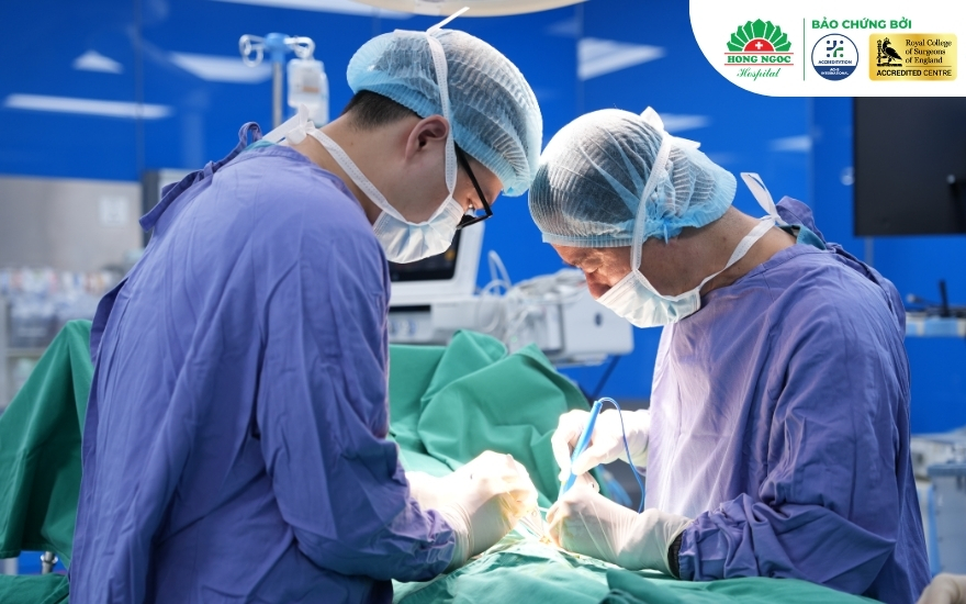
Safe and effective treatment for lung tumours at Hong Ngoc General Hospital
When facing a lung condition, choosing a reputable healthcare provider is an important part of the treatment journey. The Department of Thoracic Surgery at Hong Ngoc General Hospital provides comprehensive diagnostic and treatment services, with patients examined, advised and operated on by experienced thoracic surgery specialists.

Hong Ngoc’s quality of care is supported by a standardised, multilayered safety system. The hospital received the highest score for surgical safety under the Vietnamese Ministry of Health’s assessment criteria and holds ACHS International accreditation for healthcare quality and patient safety. It is also recognised by the Royal College of Surgeons of England as an international surgical training centre. Its operating theatres comply with UK HBN standards and apply rigorous infection control measures.
The Department of Thoracic Surgery also applies the Enhanced Recovery After Surgery programme, known as ERAS. By combining multimodal pain management, nutritional optimisation and supported mobilisation within 6 to 24 hours after surgery, the programme can help shorten hospital stays, reduce treatment costs and support an earlier return to everyday activities.
Frequently asked questions about benign lung tumours
The following medical information can help patients better understand the condition and take an active role in monitoring and treatment.
Can a benign lung tumour become cancerous?
Most true benign lung tumours have an extremely low risk of becoming cancerous. Their cells generally grow slowly, do not invade surrounding tissues and do not spread to other organs.
However, patients should continue regular monitoring as advised by their doctor. In rare cases, early malignant lesions may resemble benign tumours on chest X rays or CT scans. Follow up assessments allow doctors to monitor changes over time and reduce the risk of overlooking a potentially malignant lesion.
Does a benign lung tumour require surgery?
Not every benign lung tumour requires surgery. If the tumour is small, causes no symptoms and remains stable over time, regular monitoring may be sufficient.
Surgery may be considered when the tumour grows, compresses surrounding structures, causes recurrent infection or cannot be confidently distinguished from a malignant lesion. Patients should consult a specialist for an individual assessment.
Can a benign lung tumour disappear on its own?
Most true benign lung tumours do not disappear spontaneously. They usually grow slowly and do not invade surrounding tissues.
Regular specialist follow up remains important to assess changes in the tumour and distinguish it from early malignant lesions that may have similar imaging features.
Can a benign lung tumour grow?
A benign lung tumour may increase in size, but it usually grows slowly and does not invade surrounding tissues. Many remain stable for years without causing complications.
Patients should nevertheless attend follow up appointments as recommended so that any unusual changes can be identified and managed promptly.
At what size does a benign lung tumour require surgery?
There is no single size threshold that determines whether surgery is required. The decision is based on a comprehensive assessment of the tumour’s growth rate, symptoms, imaging characteristics and the ability to exclude malignancy.
A consultation with a thoracic surgeon is necessary to determine whether surgery or continued monitoring is the most appropriate option.
How can doctors determine whether a lung tumour is benign or malignant?
Patients cannot reliably distinguish a benign tumour from a malignant one based on symptoms alone. Doctors assess several factors, including individual risk factors, imaging characteristics and, when required, histopathological findings.
This evaluation is essential for determining whether the lesion should be monitored or treated.
Can a benign lung tumour cause haemoptysis?
Haemoptysis is uncommon in benign lung tumours but may occur because of local irritation, airway obstruction, inflammation or bleeding from a vascular lesion.
Coughing up blood can also be a warning sign of other serious respiratory conditions. Anyone experiencing this symptom should seek prompt medical assessment to identify the cause and receive appropriate treatment.
Although benign lung tumours generally do not pose the same immediate threat as lung cancer, they should not be ignored. Regular medical assessment and imaging surveillance can help identify complications such as airway compression, recurrent infection or bleeding.
Patients experiencing abnormal symptoms should seek assessment at a reputable medical facility. At the Department of Thoracic Surgery at Hong Ngoc General Hospital, specialists provide diagnosis and recommend an appropriate monitoring or treatment plan based on each patient’s condition.
—-----------------------------------------
Department of Thoracic Surgery, Hong Ngoc General Hospital
🏆 Accredited by ACHS International for healthcare quality and patient safety 🏆 Recognised by the Royal College of Surgeons of England as an international surgical training centre
Addresses:
- Hong Ngoc Phuc Truong Minh General Hospital: 8 Chau Van Liem Street, Tu Liem Ward, Hanoi
- Hong Ngoc General Hospital: 55 Yen Ninh Street, Ba Dinh Ward, Hanoi
Hotlines: 0912 002 131 / 0949 646 556
Medical disclaimer: The information provided in this article is for general reference only and does not replace medical diagnosis or treatment. Patients should visit a healthcare facility for examination, an accurate diagnosis and an appropriate treatment plan.